Since its invention back in the 16th century, the optical microscope has been widely used in biological/biomedical research and clinics to analyze cells and tissues. In general, an optical microscope uses white light from a light bulb or fluorescence from staining dyes to visualize cell/tissue structures and morphologies, which we call bright-field microscopy and fluorescence microscopy. Although these microscopes are widely used for diagnosing cancer, cancer or cancer precursor may be in progress already at the biomolecular level (e.g., at the DNA level), but the structural and morphological changes (such as appearance of polyps or emergence of actual tumors), which occur at a much later stage in cancer progression, are still invisible to traditional microscopy. These traditional microscopes are not able for early cancer diagnosis.
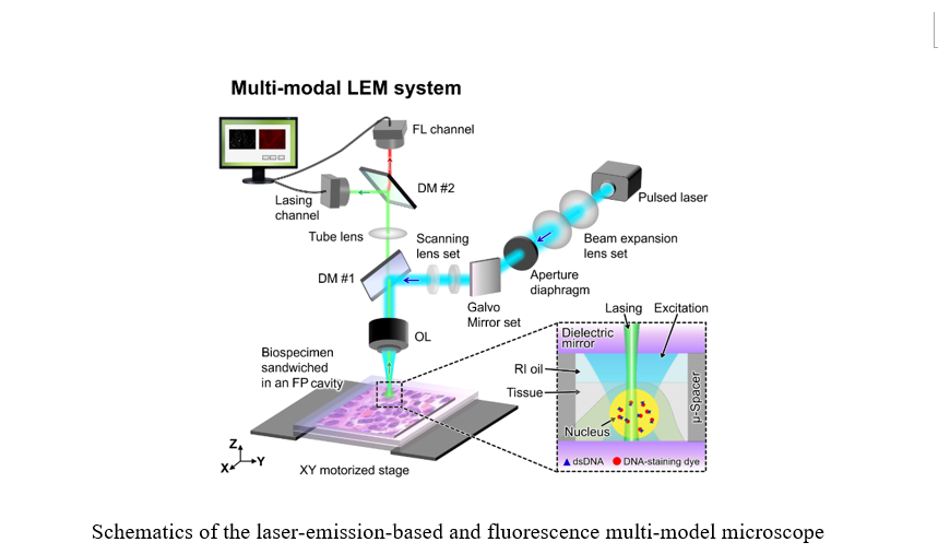
In this project, the team conceptualized and developed a new type of microscopy – laser emission-based microscopy (LEM), in which cells and tissues stained with dyes are made to lase, and the laser emission, rather than fluorescence, is used to analyze cells/tissues Laser emission has unique advantages over fluorescence, including narrow linewidth, threshold behavior, and strong intensity, leading to ultrasensitive detection, superior image contrast, and higher spectral/spatial resolution.
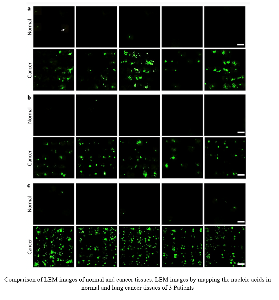
Recently, LEM was explored to examine Stage I/II lung cancer tissues from human patients, as well as human colon/stomach/breast tissues. We found that the cancer cells/tissues consistently have a much lower lasing threshold than the normal cells/tissues when their nuclei are stained with dyes.
Even more, our recent studies on dietary intervened mice have confirmed the LEM’s capability to detect abnormal DNA/chromatin activities in the “apparently normal” colon tissues from the mice at risk for colon cancer even there is no observable polyps, lesions, or tumors in those tissues.
Currently, the global histology and cytology market has reached more than 14 billion dollars. The LEM is an emerging imaging technology that may provide a new tool that complements the traditional H&E and IHC methods for early cancer diagnosis/prognosis, as well as intra-operative tumor margin detection. It will expand the market size even more.
Now, a multi-modal LEM system development and clinical trials are ongoing.